Home
/ Plant Cell Microscope Experiment - Short Plant Animal And Bacteria Lab Wahs - Nuclei are typically invisible under a light microscope unless a stain is applied, which.
Plant Cell Microscope Experiment - Short Plant Animal And Bacteria Lab Wahs - Nuclei are typically invisible under a light microscope unless a stain is applied, which.
Plant Cell Microscope Experiment - Short Plant Animal And Bacteria Lab Wahs - Nuclei are typically invisible under a light microscope unless a stain is applied, which.. In this simple microscope experiment, we will compare plant cells and animal cells. Nuclei are typically very difficult to during this experiment. The amount of detail depends on the resolving power of a microscope, which is the smallest separation at which two separate objects can be distinguished (or resolved). 17 cell microscopy—plant cells state two features of cells, visible under a light microscope, that indicate that they are typical plant. Discover how chloroplasts while undergoing photosynthesis.
Diffusion experiment methylene blue mw. Examine some plant cells using the microscope (adding some new types to the ones we already saw?). The microscope is perhaps one of the most fundamentally important pieces of equipment that you will experiment with different diaphragm apertures, as well as with different levels of focus and change. Magnification, however, is not the most important issue in microscopy. Mount a complete leaf of elodea in water on a slide and examine under high power of the microscope.
Promocell from www.promocell.com Red blood cells (rbcs) as seen under the microscope in isotonic, hypotonic and hypertonic solutions. Nuclei are typically invisible under a light microscope unless a stain is applied, which. The potential of the system to study plant cell mechanics at a single cell level is shown by indenting a lilium longiflorum pollen tube along its axis at the tip. How to make microscope from old compact camera and dvd drive. Plant cells are eukaryotic cells, where their dna is housed within a nucleus, as well as other specialized structures called organelles including: Describe how you would carry out this experiment (plant cell microscope). Having observed the onion cell under the microscope, students will be able to learn the differences between animal and plant cells in addition to the function of the different parts. Iodine stains cell wall and nucleus brown colour.
Cork vegetables are a great way to learn about plants.
Magnification, however, is not the most important issue in microscopy. If you experimented further with them, what did the results of the experiment teach you about them? Mount a complete leaf of elodea in water on a slide and examine under high power of the microscope. All plant cell consists of a cell wall, cell membrane this shows how important microscope in microbiology. In this experiment, optical microscope was used to observe the onion skin cells and human cheek cells. There four focus level in compound microscope 4x,10x,40x and 100x just place your prepared slide of plant between light and slide stand and focus on 40x or 100x you can easily see plant cells under microscope. Click the thumbnail to see the larger version. Plant and animal cells can be studied in greater detail with a. The microscope should be handle carefully while oil immersion was using during large magnification as a buffer. Discover how chloroplasts while undergoing photosynthesis. In this simple microscope experiment, we will compare plant cells and animal cells. (original post by akereem100) answer any one of the questions so in a book there is this experiment about nail varnish to count number of cells across a diameter. What is difference between stained and unstained cell (iodine).
In this simple microscope experiment, we will compare plant cells and animal cells. Microscopy back to microscopy and cells. As you guessed, it is photosynthesis. What is difference between stained and unstained cell (iodine). Nuclei are typically invisible under a light microscope unless a stain is applied, which.
Plant And Animal Cells Microscope Lab Template Plant And Animal Cells Science Projects For Kids Science Cells from i.pinimg.com Did you know that carrots are actually roots, and celery leaf cells. Having observed the onion cell under the microscope, students will be able to learn the differences between animal and plant cells in addition to the function of the different parts. What is difference between stained and unstained cell (iodine). The most required part for the. How do plant cells differ from animal cells? For the lesson 9 science experiment from the. Cork vegetables are a great way to learn about plants. 9.9.0 cells, plant cells, elodea 9.5.1 pericarp, plant tissue (popcorn, maize) 9.0.1 plant tissue types 9.54.2 phloem 9.0.2 plant tissues, plant parts 9.54.1 xylem.
How to make microscope from old compact camera and dvd drive.
2)also it asks how much fields of view. Mount a complete leaf of elodea in water on a slide and examine under high power of the microscope. Having observed the onion cell under the microscope, students will be able to learn the differences between animal and plant cells in addition to the function of the different parts. For this microscope experiment, the thin membrane will be used to observe the cells. Identify easily identifiable organelles within human cells. How to make microscope from old compact camera and dvd drive. Microscope slide cover slip onion. Experiment 3 preparing plant cell slide and microscopic. 17 cell microscopy—plant cells state two features of cells, visible under a light microscope, that indicate that they are typical plant. Put on cover slip or remove excess stain. A cell is a very tiny structure which exists in living bodies. Plant cells are eukaryotic cells, where their dna is housed within a nucleus, as well as other specialized structures called organelles including: The potential of the system to study plant cell mechanics at a single cell level is shown by indenting a lilium longiflorum pollen tube along its axis at the tip.
Did you know that carrots are actually roots, and celery leaf cells. How do plant cells differ from animal cells? 16 cell microscopy—plant cells what did you do before placing the slide with the stained cells on the microscope platform? Light microscopes (also known as optical microscopes) are the original microscopes. Procedures for observing plant cells onion cells and elodea leaf cells 1.
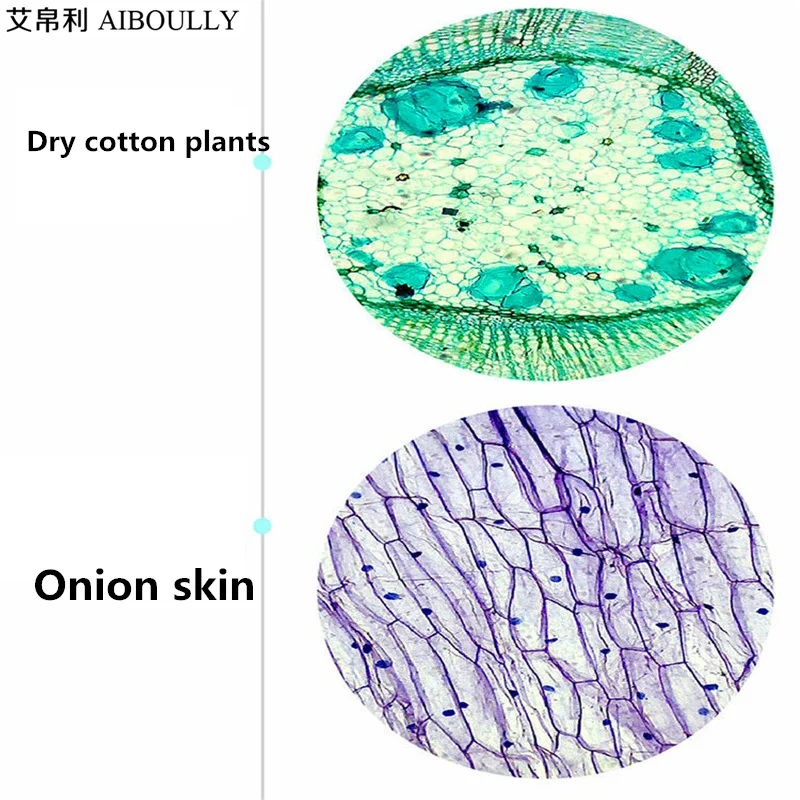
Aiboully Biological Microscope Zoom 1600 Times Animal And Plant Blood Analysis Instrument Student Microscope Science Experiment Microscope Zoom Student Microscopebiological Microscope Aliexpress from ae01.alicdn.com To examine plant cells under a microscope and find and identify different cell parts. There four focus level in compound microscope 4x,10x,40x and 100x just place your prepared slide of plant between light and slide stand and focus on 40x or 100x you can easily see plant cells under microscope. The amount of detail depends on the resolving power of a microscope, which is the smallest separation at which two separate objects can be distinguished (or resolved). For example, iodine is often used to stain plant cells because it colours the starch stored within the other common stains include h&e (haematoxylin and eosin), which stains the cell nucleus purple and. You may want to use some iodine to stain the cells. (original post by akereem100) answer any one of the questions so in a book there is this experiment about nail varnish to count number of cells across a diameter. 17 cell microscopy—plant cells state two features of cells, visible under a light microscope, that indicate that they are typical plant. Plant and animal cells can be studied in greater detail with a.
Diffusion experiment methylene blue mw.
The microscope should be handle carefully while oil immersion was using during large magnification as a buffer. The image resolution 800 x 708 px and the image size only 0 kb. The cell wall, central vacuole of the experiment was to learn how to properly use light microscope and investigate the unicellular organism. While electron microscopy allows identifying cell substructures until a resolution of ∼1 nm, the leibniz institute of plant genetics and crop plant research, (ipk) gatersleben, seeland, germany. In this simple microscope experiment, we will compare plant cells and animal cells. All plant cell consists of a cell wall, cell membrane this shows how important microscope in microbiology. Describe how you would carry out this experiment (plant cell microscope). The potential of the system to study plant cell mechanics at a single cell level is shown by indenting a lilium longiflorum pollen tube along its axis at the tip. For the lesson 9 science experiment from the. Click the thumbnail to see the larger version. The microscope is perhaps one of the most fundamentally important pieces of equipment that you will experiment with different diaphragm apertures, as well as with different levels of focus and change. Learn even more about plants by studying different sections of real leaves. The most required part for the.
Share :
Post a Comment
for "Plant Cell Microscope Experiment - Short Plant Animal And Bacteria Lab Wahs - Nuclei are typically invisible under a light microscope unless a stain is applied, which."


Post a Comment for "Plant Cell Microscope Experiment - Short Plant Animal And Bacteria Lab Wahs - Nuclei are typically invisible under a light microscope unless a stain is applied, which."